or
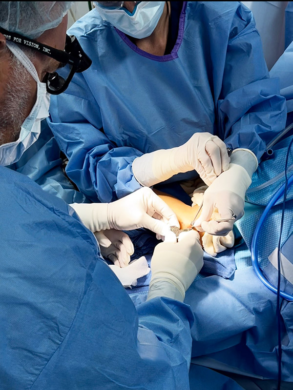
Guide placement – Photo 01
Dr. Badia is seen placing the guide, which will allow him to visualize the ulnar nerve as well as the ligament that will be divided.
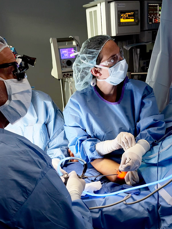
Endoscopic Cubital Tunnel Release – Photo 03
Dr. Badia uses an endoscope to look into the cubital tunnel, where the ulnar nerve has just been released.
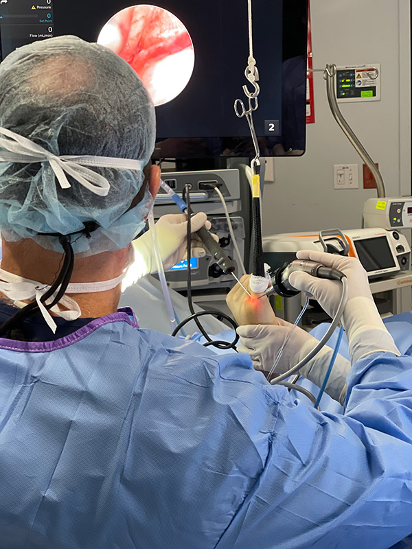
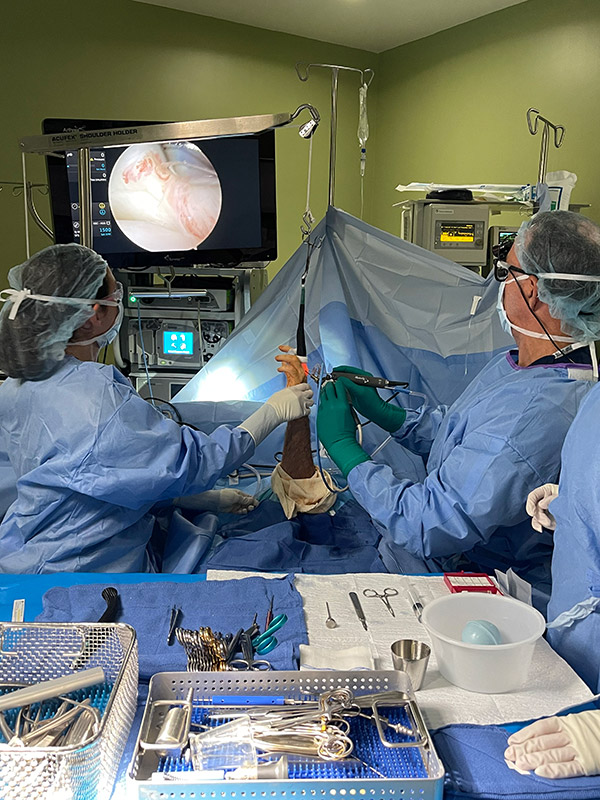
Metacopophalangeal Joint Arthroscopy – Photo 01
Metacopophalangeal Joint Arthroscopy to Treat an Articular Fracture
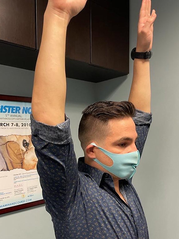
Patient’s motion after clavicle ORIF – Photo 03
This patient regained full motion only weeks after clavicle ORIF
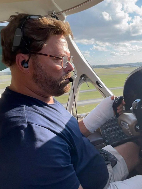
Patient flies himself home after surgery – Photo 07
Our patient felt so well after surgery, he flew himself home! Not to worry, his co-pilot was ready to take over if needed.
